
Standard X-rays show a two-dimensional view. Cone beam imaging creates a 3D model, allowing accurate measurements and views from every angle, which helps with complex cases.
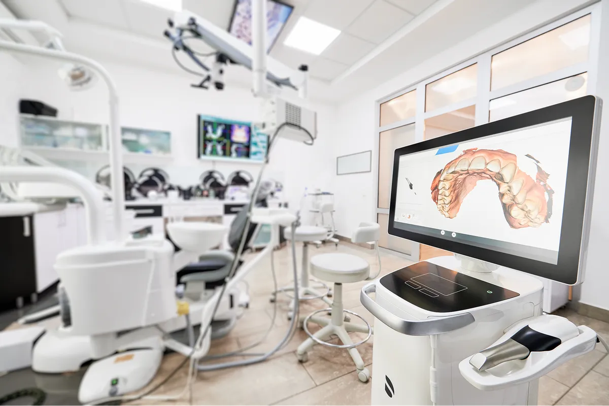
Cone Beam Imaging
Cone Beam Imaging in Mt. Prospect, Elk Grove Village, Bartlett and Chicago, IL: Your 3D Dental X-Ray Guide
Cone beam imaging offers a precise, three-dimensional view of your teeth, jaws, and facial structures. At Associates For Oral, Maxillofacial & Implant Surgery, LTD, this technology supports accurate diagnosis and treatment planning for oral surgery,
Cone beam imaging offers a precise, three-dimensional view of your teeth, jaws, and facial structures. At Associates For Oral, Maxillofacial & Implant Surgery, LTD, this technology supports accurate diagnosis and treatment planning for oral surgery, dental implants, impacted teeth, and more. Patients can expect clear images and a comfortable, efficient scan experience through the use of cone beam imaging in Mt. Prospect, Elk Grove Village, Bartlett and Chicago, Illinois.
Cone Beam Imaging Explained
Cone beam computed tomography (CBCT) is a 3D dental X-ray that captures detailed images of bone, teeth, nerves, sinuses, and airway. Unlike traditional two-dimensional X-rays, CBCT provides a volumetric image that can be viewed from multiple angles. This makes it easier to see exact tooth positions, bone quality, and anatomical variations.
CBCT differs from a hospital-based medical CT scan by using a cone-shaped X-ray beam and a single rotation around your head, which generally results in lower radiation than a conventional medical CT. It delivers more information than panoramic or bitewing X-rays, which is why it is often used for complex dental decisions such as implant placement or evaluating impacted canines.
Benefits of Cone Beam Imaging
- Enhanced diagnostic detail for teeth, bone, nerves, and sinuses.
- Accurate implant planning with measurements of bone height and width.
- Improved evaluation of impacted teeth and root anatomy.
- Clear visualization for jaw joint, airway, and sinus assessments.
- Targeted treatment with fewer surprises during procedures.
- Efficient planning that can reduce the number of appointments.
How Cone Beam Imaging Works
The scan process is brief and straightforward. The machine rotates around your head while a cone-shaped beam takes multiple images, which software reconstructs into a 3D model. Here is a typical step-by-step guide:
- Preparation: You will remove glasses, earrings, dentures, or metal objects that could affect the image.
- Positioning: You will sit or stand while your head is gently stabilized.
- Scanning: The unit rotates for about 10 to 20 seconds, capturing the data.
- Processing: Images are reconstructed into a 3D view for precise measurements.
- Review: Your provider reviews the scan to discuss findings and next steps.
Your Scan Experience: What To Expect
Most CBCT scans take only a few minutes from start to finish. The scan is painless and does not require anesthesia. You will hear a soft humming sound while the machine rotates. Breathing normally and staying still help produce a crisp image.
Radiation exposure with dental CBCT is typically higher than a panoramic X-ray but lower than a traditional medical CT scan. Practices follow the ALARA principle—keeping radiation “as low as reasonably achievable”—and use settings appropriate for diagnostic needs. If you are pregnant, think you might be pregnant, or have specific health concerns, tell the team before imaging so alternatives or additional precautions can be considered.
This technology is often used for dental implants, root canal retreatment, bone graft planning, wisdom tooth assessment, and evaluating the temporomandibular joint. Patients seeking what is cone beam imaging, how does CBCT work, and CBCT vs CT scan comparisons will find that the 3D view provides the detail needed to guide safe, effective care.
For questions about cone beam imaging in Mt. Prospect, Elk Grove Village, Bartlett and Chicago, IL, or to arrange a scan requested by our oral surgeons, contact Associates For Oral, Maxillofacial & Implant Surgery, LTD today.

Cone Beam Imaging
Ready to Get Started?
Our experienced team is here to guide you through every step of your treatment.
Contact us today to schedule your consultation and take the first step toward better oral health.
Frequently Asked Questions About Cone Beam Imaging
Yes. CBCT uses X-rays, so there is radiation exposure, but it is generally lower than a medical CT. Imaging is ordered only when the benefits outweigh the risks.
The actual scan usually takes 10 to 20 seconds. Expect a short visit to allow for positioning, image processing, and review of the results.
The scan is painless, and the unit is open on all sides. Most people do not feel confined. Staying still is the main request during imaging.
CBCT helps evaluate bone quality for implants, locate nerves and sinuses, assess impacted teeth, analyze root canals, and plan surgeries with precision.
Remove metal accessories and let the team know if you are pregnant. No fasting or special medication is typically required.
Yes. Digital files can be securely shared with your dental providers to coordinate care and support treatment planning.

Contact Us Today
Schedule Your Consultation